Sie befinden sich hier
Inhalt
Our clinical research addresses unresolved problems in vascular medicine by combining deeply characterized patient cohorts, advanced vascular imaging, biomarkers and prospective clinical studies. We aim to understand why vascular disease develops and recurs, identify patients at risk and translate mechanistic insights into better diagnosis, prevention and treatment.
1. Blood Flow, Vascular Disease and Precision Ultrasound
Why do some arteries develop recurrent disease after apparently successful revascularization?
Clinical Problem
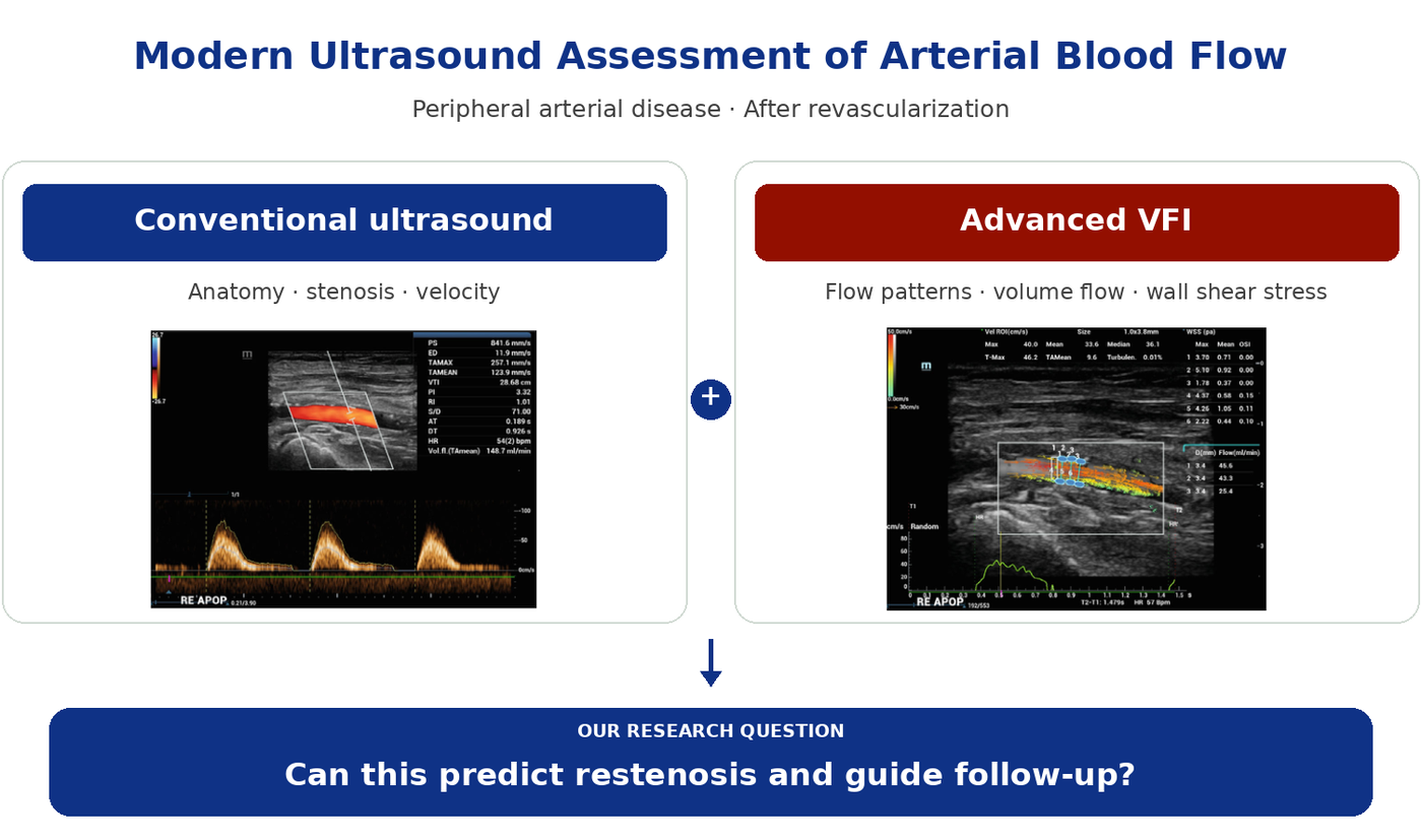
Patients with peripheral arterial disease frequently require revascularization to restore blood flow. However, successful revascularization does not always result in durable vascular recovery, and restenosis remains an important clinical problem. Conventional vascular ultrasound primarily assesses stenosis and blood-flow velocities but provides limited information about the local hemodynamic forces acting on the vessel wall.
Our Approach
We combine prospective clinical studies in patients with peripheral arterial disease with conventional duplex ultrasound and high-frame-rate vector flow imaging (VFI). We characterize blood-flow direction and velocity, volume flow, wall shear stress and oscillatory shear index before and after vascular interventions.
What We Have Already Shown
Our initial work demonstrated the feasibility of VFI in the femoropopliteal arteries of patients with PAD. We subsequently demonstrated measurable changes in blood volume flow and wall shear stress following iliac or femoropopliteal revascularization.
Next Steps & Clinical Impact
We now want to determine whether these hemodynamic parameters can help predict restenosis, identify patients who may benefit from closer surveillance and ultimately enable more individualized treatment monitoring.
Key Publications
J Ultrasound Med. 2026
Prüser JL, Winkelbauer G, Dieker W, Adler A, Amendt K, Sigl M. Pre- and Post-Interventional Hemodynamic Characterization of the Femoropopliteal Artery Using Vector Flow Imaging in Peripheral Arterial Occlusive Disease. J Ultrasound Med. 2026;45:1397–1407. doi:10.1002/jum.70176.
Vasa. 2023
Shchetynska-Marinova T, Gerdes L, Hohneck AL, Winter L, Amendt K, Schwenke K, Gerken ALH, Du Y, Dürschmied D, Sigl M. First experiences of ultrasound vector flow imaging at the femoropopliteal artery in peripheral arterial disease. Vasa. 2023;52:394–401. doi:10.1024/0301-1526/a001095.
2. Aneurysm Disease: Screening, Imaging, and Perioperative Risk Assessment
How can we detect aneurysms earlier, identify patients at risk, and make treatment safer?
Clinical Problem
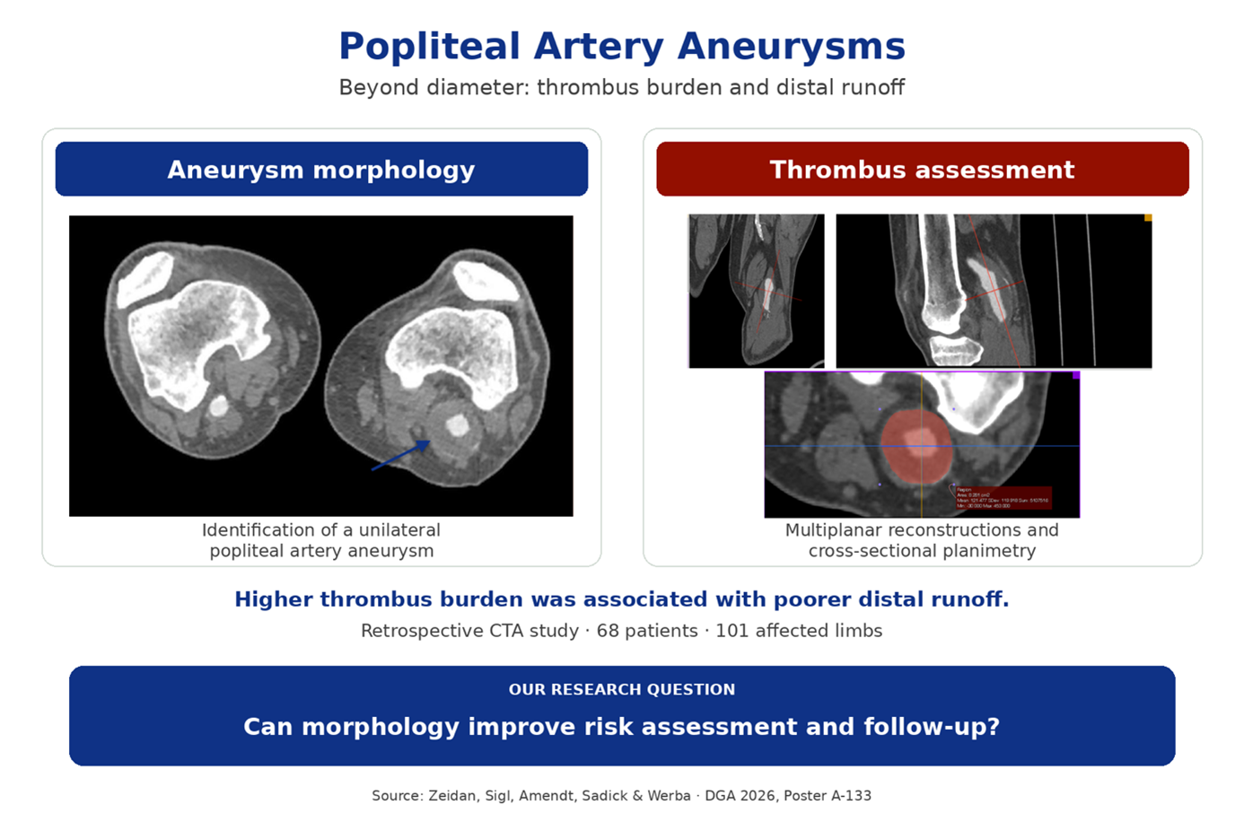
Aneurysms may remain undetected until rupture, thrombosis, or embolisation occurs. Early diagnosis and reliable risk assessment are essential, but aneurysm diameter alone does not fully describe disease severity. Cardiovascular comorbidities also influence the safety of treatment.
Our Approach
We combine targeted ultrasound screening, detailed vascular imaging, and structured cardiovascular assessment. Our clinical studies and systematic reviews examine aneurysm morphology, intraluminal thrombus burden, distal runoff, and surveillance after repair, alongside perioperative outcomes following open aortic surgery.
Next Steps & Clinical Impact
We aim to integrate prospectively collected clinical, laboratory, and imaging data to identify risk factors and blood biomarkers associated with the presence and size of aneurysms. These findings could help refine targeted screening and improve individualised risk assessment and follow-up.
What We Have Already Shown
Our screening studies identified previously unrecognised aortic and peripheral aneurysms in high-risk patients. Our systematic reviews highlighted inconsistent definitions and measurement methods for popliteal aneurysms and limited evidence for contrast-enhanced ultrasound surveillance after repair.
In our recent CTA study, greater thrombus burden was independently associated with impaired distal runoff, whereas maximum aneurysm diameter was not. Our work also linked structured preoperative cardiac assessment to fewer early cardiovascular complications after open abdominal aortic surgery.
Key Publications
Publications
Zeidan Y, Sigl M, Amendt K, Sadick M, Werba N. Intraluminal thrombus burden is an independent imaging predictor of distal runoff impairment in popliteal artery aneurysms. Vasc Med. 2026. Published online October 7. doi:10.1177/1358863X261478067.
Keller S, Zeidan Y, Sigl J, Dieker W, Naumer L, Amendt K, Sigl M. Contrast-enhanced Sonography for Detecting Endoleaks After Popliteal Artery Aneurysm Repair: A Systematic Review. In Vivo. 2026;40:704–714. doi:10.21873/invivo.14232.
Zeidan Y, Sigl J, Dieker W, Gerken ALH, Gerdes L, Sigl M. Defining Popliteal Artery Aneurysm: A Systematic Review of the Morphological Assessment Used for Diagnosis and Treatment. Ann Vasc Surg. 2025;120:205–222. doi:10.1016/j.avsg.2025.06.012.
Sigl M, Baumann S, Könemann AS, Keese M, Schwenke K, Gerken ALH, Dürschmied D, Rosenkaimer S. Prognostic value of extended cardiac risk assessment before elective open abdominal aortic surgery. Herz. 2024;49:210–218. doi:10.1007/s00059-023-05209-y.
Hohneck A, Shchetynska-Marinova T, Ruemenapf G, Pancheva M, Hofheinz R, Boda-Heggemann J, Sperk E, Riffel P, Michels J, Borggrefe M, Akin I, Sigl M. Coprevalence and Incidence of Lung Cancer in Patients Screened for Abdominal Aortic Aneurysm. Anticancer Res. 2020;40:4137–4145. doi:10.21873/anticanres.14413.
Hohneck A, Keese M, Ruemenapf G, Amendt K, Muertz H, Janda K, Akin I, Borggrefe M, Sigl M. Prevalence of abdominal aortic aneurysm and associated lower extremity artery aneurysm in men hospitalized for suspected or known cardiopulmonary disease. BMC Cardiovasc Disord. 2019;19:284. doi:10.1186/s12872-019-1265-2.